With high temporal and spatial resolution retinal motion tracking and concurrent cell-resolved imaging, we can analyze the cellular pattern of cone activation during psychophysical interrogation. How does microscopic eye motion modulate visual perception? These and other questions can be answered by using the AOSLO and bSLO as high-speed retinal tracker with the capability to also manipulate the retinal slip of a visual stimulus with cellular accuracy.
Kavitha Ratnam, Niklas Domdei, Wolf Harmening*, Austin Roorda* (2017)
Benefits of retinal image motion at the limits of spatial vision.
Further Reading
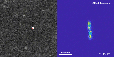
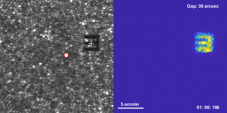
A small visible stimulus projected on the retina will move across many foveal photoreceptors during fixation. The retina was stabilized to better show the relative trajectory of the stimulus. In reality, the stimulus was immobile, and the retina moved. Together with a high resolution map of the cone photoreceptor mosaic, such trajectories can be used to compute the exact spatio-temporal activation pattern for each cone.
The white circle is the centroid of highest cone density. Individual presentations, each 0.5 sec in duration, were concatenated for this video. Both videos show one 20-trial experimental run to determine a discrimination acuity threshold (top) or positional hyperacuity threshold (bottom), with the current estimate given in arcsec.
Timecode is Trial:Frame:ms
Veronika Lukyanova, Jenny Reiniger, Bilge Sayim, Wolf Harmening (2021)
Do fixational eye movements help or hinder hyperacuity.
Poster at Neurocon, 5. Nov. 2021, Günzburg, Germany