Further reading
Domdei N, Reiniger JL, Pfau M, Charbel Issa P, Holz FG, Harmening WM (2016) Histology in the living eye: non-invasive microscopic structure and function analysis of the retina with adaptive optics.
Der Ophthalmologe, 114(3): 206-214 [doi:10.1007/s00347-016-0411-9]
Harmening WM, Sincich LC (2019)
Adaptive Optics for Photoreceptor-Targeted Psychophysics.
In: Bille J (ed.) High resolution Imaging in Microscopy and Ophthalmology, Springer, Cham. pp 359-375
[doi:10.1007/978-3-030-16638-0_17]
For German readers:
Reiniger JL*, Domdei N*, Pfau M, Müller PL, Holz FG, Harmening WM (2017) Potential of adaptive optics for the diagnostic evaluation of hereditary retinal diseases.
Klinische Monatsblätter der Augenheilkunde, 234: 311-319.
[doi:10.1055/s-0043-100631]
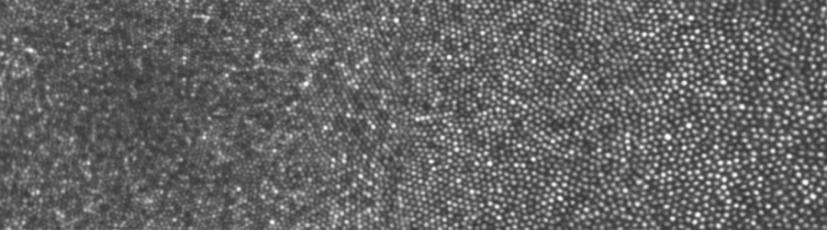
With the tools in the lab we are able to image the healthy and diseased eye with cellular resolution, revealing even the smallest cones and rod photoreceptors in the living retina. We aim to improve our microstimulation techniques to a degree where it can be of advantage for clinically relevant experimentation.
Click the icons to download our PubMed referenced directory of retinal pathologies studied with adaptive optics in XLS or PDF format (last update Sept. 2021, compiled by Niklas Domdei, Maximilian Pfau and Julius Ameln).